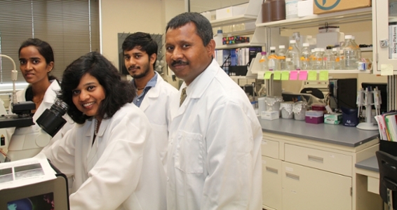
Dr. Narendra Kumar's Lab
Research Focus

Obesity-associated metabolic syndrome (MetS) is both the US and a worldwide epidemic and a major burden to the healthcare system. Chronic low-grade inflammation (CLGI) is a well-established characteristic of the obese-human condition and conventionally, research has focused on the CLGI of the liver and adipose tissue as a driver. Though, the gastrointestinal (GI) mucosa is the first tissue that interacts with dietary components and luminal microbiota both of which are known to regulate obesity, the research on the role of GI-mucosa in obesity-associated MetS has been ignored. Recent novel findings from my lab support a key role of Janus kinase 3 (Jak3), a non-receptor tyrosine kinase, in intestinal and systemic CLGI associated obesity and diabetes in both an animal model and in humans. Overall, the existing literature, our publications, and additional unpublished data indicate that Jak3 regulates; colonic and systemic CLGI, and multiple symptoms of basal- and diet-induced obesity and diabetes including, glucose regulatory phenotype, hyperinsulinemia, and liver steatosis. Although the combined evidence supports an essential role for Jak3 in CLGI, a characteristic precursor for obesity and diabetes, there is a critical need to determine the associated underlying mechanisms. In the absence of such knowledge, the potential to identify intestinal-specific factors regulating obesity and diabetes and develop intestinal-based therapeutic interventions to inhibit CLGI characteristic of diabetes will likely remain limited. The research focus of our lab is to determine tissue-specific roles of Jak3 and associated signaling complexes in CLGI-onset as a precursor of multiple chronic diseases including obesity, diabetes, Alzheimer, and inflammatory bowel disease with specific goals to determine a timely intervention to prevent aforementioned complications.
Inflammatory bowel disease (IBD) that includes Crohn’s disease and Ulcerative colitis is a chronic inflammatory condition of the gastrointestinal tract. Though the annual death from these diseases is over 50,000.00, the incidences of new cases have been rising to be over 100,000 annually. The intestinal epithelial wound repair functions (Restitution) are compromised during various intestinal disorders including IBD. The research focus of our lab is to dissect the roles of intestinal epithelial, intestinal immune cells and gut microbiota in mucosal restitution and formation of barrier functions. Our lab was first to report the functions of Jak3 in intestinal epithelial mucosa. Our studies show that in an intestinal epithelial cells IL-2 (a cytokine produced during intestinal inflammation) promotes mucosal wound repair where activated Jak3 forms complex with villin and ShcA in an IL-2 dependent manner. We have also shown that that either inhibition of Jak3 activation or intestinal mucosal tissue-specific knock out of Jak3 results in loss of tyrosine phosphorylation of villin, p52ShcA, and beta-catenin, and a significant decrease in mucosal wound repair. Our hypothesis is that JAK3-mediated signaling plays an essential role in intestinal epithelial restitution and barrier functions during normal physiological condition and during intestinal inflammation. Studies are underway to define the tissue-specific Jak3-mediated signaling pathways that regulate CLGI as a precursor for the development of not only IBD but also associated various complication including neurodegeneration and Alzheimer disease.
Protocols
Prepare Frozen Tissue Sections
- Place a freshly dissected tissue block (<5mm thickness) onto a pre-labeled tissue base mold.
- Cover the entire tissue block with cryo-embedding media (e.g OCT)
- Place the base molds containing the tissue block into liquid nitrogen and freeze completely.
- Store the frozen tissue block at -80 οC until ready for sectioning.
- Transfer the frozen tissue block to a cryotome cryostat at -20 οC and allow the temperature to equilibrate to the temperature of the cryotome.
- Section frozen tissue block into the desired thickness (typically 5 μm) using the cryotome and place the tissue sections onto superfrost glass slides.
- Dry the sections at room temperature overnight or store in -80οC for later use.
Immunofluorescence Staining of Frozen Tissue Sections
- Fix the tissue sections in 300 ml of 4% PFA taken in coplin jar for 5 min.
- Rinse the slides in phosphate-buffered saline at neutral pH for 3 changes in 3 different coplin jars, 5 mins each.
- Incubate the slides in 1% BSA in Dulbecco’s Phosphate-Buffered Saline (DPBS) containing 0.1% Triton-X-100 for 30 mins, at room temperature.
- Drain off the blocking buffer and Make liquid blocker around the tissue and allow drying for 15 sec.
- Apply 100μl of an appropriately diluted primary antibody in 1% BSA in DPBS containing 0.1% Triton-X-100 to the sections on the slides and incubate the slides in a humidified chamber for 1h at room temperature or overnight at 4οC.
- Rinse the slides in 300 ml DPBS for 3 changes in 3 different coplin jars, 5 mins each.
- Apply 100μl of an appropriately diluted secondary antibody in 1% BSA in DPBS containing 0.1% Triton-X-100 to the sections on the slides and incubate the slides in a humidified chamber for 1h at room temperature.
- Rinse the slides in 300 ml DPBS for 3 changes in 3 different coplin jars, 5 mins each.
- Mount the coverslips gently onto the slides using 20μl Vectashield Mounting Medium containing DAPI for nuclear staining.
Staining Cell Surface Antigens
- Aliquot 100μL of cells per tube (1x10⁶ cells total). Add 20 μL blocking reagent containing 2.4 G2 (anti- Fc receptor (0.5-1μg) for mouse cells. Incubate 20 mins on ice.
- Add appropriately diluted unconjugated primary antibodies to each tube, vortex and incubate for 60 mins at 4ο C. Avoid exposure to light.
- Add 200μL of 1x PBS to each tube to wash off excess antibody. Centrifuge for 5 mins at 250xg. Aspirate the supernatant, being careful not to disturb the pellet.
- Add 100μL of 1X PBS to each tube. Add appropriately diluted fluorochrome-conjugated secondary antibodies to tubes. Vortex and incubate for 30 mins at 4οC. Avoid exposure to light.
- Add 2 ml of 1x PBS to each tube to wash off excess antibody. Centrifuge for 5 mins at 250xg. Aspirate the supernatant, being careful not to disturb the pellet.
- [Optional] Add a viability dye; either 7-AAD or Propidium iodide (5 μl/sample) to exclude dead cells from analysis.
Staining Intracellular Antigens
- Stain cell surface antigens as described above (step 1 to 6)
- After the last wash, discard the supernatant and pulse-vortex the sample to completely dissociate the pellet.
Fixation and Permeabilization
Fixation in 0.01% formaldehyde. Permeabilization in Triton-X-100 or NP-40 (0.5% in PBS)
- Add 100 μL of fixative and incubate for 10 mins at room temperature.
- Add 100 μL detergent based permeabilizing agent and incubate at room temperature for 15 minutes.
- Centrifuge the cells at 300xg for 5 mins, discard the supernatant and re-suspend the pellet in remaining volume.
- Follow antibody staining protocol as indicated in our “direct” and “indirect” protocols.
Note: If combining both direct and indirect staining together, the sequence of staining will be: Primary antibody followed by fluorochrome-conjugated secondary antibody and then stain directly (fluorochrome-conjugated primary antibody).
- Weigh the tissue sample in a 50 mL tube.
- Keep the sample on ice and wash with ice-cold 1x PBS and aspirate off the PBS.
- Repeat until wash buffer appears clear.
- Add sufficient volume of cold lysis buffer with 1X PIC, enough to cover the sample. Note: RNase/ Dnase (0.02 mg/ml) may be added to the samples to facilitate RNA and DNA digestion.
- Grind/homogenize tissue in a tube and incubate on ice for up to 60 mins.
- Transfer mixture to microcentrifuge tubes and spin at 14, 00 rpm for 40 mins at 4οC.
- Poke through lipid layer and remove supernatant. Discard the cellular debris and lipids.
- If necessary, re-spin supernatant at 14, 000 rpm and repeat step 7 to obtain clean lysate free of debris and lipids.
- Determine the protein concentration using the Bradford Protein Assay.
- Combine equal volumes of 2X SDS sample buffer and cell lysate supernatant. Pipet up and down or vortex several times to mix. Aliquots can be stored at -80οC for further use. Prior to use, heat samples at 95-100οC for 3-5 mins and then load immediately on an SDS-PAGE gel. Avoid freeze/thawing lysate as much as possible.
1x Lysis Buffer Composition
- 10ml\A Tris pH 8.0 – 1.6 mg/ml Benzamidine HCL
- 130 ml\A NaCl -1.0mg/ml Phenanthroline
- 1% Triton-X-100 – 1.0 mg/mL Aprotonin
- 10 ml\A NaF – 1.0 mg/ml Leupeptin
- 10 ml\A NaPi pH 7.5 (sodium phosphate)- 1.0 mg/ml Pepstatin A
- Collect background blood sample (~75 μL; to have at least 12 μL of plasma) from the mice into tubes containing EDTA.
- Inject 3 μg deuterium oxide/g body weight intraperitoneally into the mice.
- After 1 hour, collect a blood sample (~75 μL; to have at least 12 μL of plasma) from the mice into tubes containing EDTA.
- Centrifuge the blood samples to obtain plasma
- Store at -80ο C
Note: Enrichment of approximately 0.2% to 0.4% is achieved at 3 μg deuterium oxide/g body weight.
Animal Experiments
Animal housing for conducting experiments using genetically modified rodents, gene delivery in live animals, animal tagging, glucose monitoring, body fat monitoring, aseptic surgery.
Bioinformatics & Other Software
Gold docking suite, Sequence scanner, Chroma, Origin, NIS element, CFlow.
Histopathology
Cryostat CM 1510 S (Leica), Leica HI1220 Flattening Table for Histology, Fully Motorized Rotary Microtome RM2255 (Leica), Leica HI1210 Water Bath, all the equipment and supplies for histochemistry & immunohistochemistry.
Mammalian Cell Culture
Dedicated cell culture facility that includes sterile CO 2 Incubator (Thermo Scientific), Laminar flow hoods (Labconco), Refrigerated Centrifuge (Thermo Scientific), Pipettes aids, Water bath (ISO temp), inverted microscope (Labomed), Aspirator (Welch), EVOM epithelial TEER Voltohmeter (World Precision Instruments), and liquid nitrogen storage.
Microscopy
Nikon Eclipse TS 100 Microscope (Nikon), Motorized stage and NIS elements AR 3.0 (Nikon), Microscope TCM 400 (Labomed)
Recombinant Protein Production & Structure-Function Analysis
Recombinant protein production and purification facility that includes incubator shaker (New Brunswick Scientific), Bacterial incubator (IB-05G), Large volume refrigerated centrifuge (Thermo Scientific), Two-block and one block PCR machine, Gel-filtration system, Isoelectric focusing system (Rotofor Cell; BioRad), -80 ͦC glycerol stock storage facility.
SDS-PAGE & Immunoblotting
Gel Electrophoresis Unit with blotting apparatus (Bio-Rad), Electrophoresis Power Pack (Thermo Fisher), Bio-Rad ChemiDoc XRS + with image lab TM software (Bio-Rad), Micro-Centrifuge (Thermo Scientific), Over Head Stirrer (Tissue Homogenizer) (Wheaton), Power Pack (Bio-Rad), Three tire Infinity Rocker (Next Advance), Plate Reader (Awareness Technology Inc).
Other Lab Facilities
Electronic Balance (Satorius), Hot Plate Stirrer (VWR), ISO temp Refrigerated Water Circulator (ISO temp), Water bath (VWR), Mini Blot Mixer (VWR), Rotofor® and Mini Rotofor Cells (BioRad), Multi Mix Rotator (VWR), Dual block PCR machine (G-storm), pH Meter (Jenco Instruments), Refrigerator -20 oC (Kenmore), Refrigerator 4 oC (Electrolux), Double door Refrigerator 4 oC (ISOTEMP), Excella E-24 Incubator Shaker (New Brunswick Scientific), UV Trans illuminator (Ultra Violet Products), Mini Pump (CONTROL), Rotaphor isoelectric focusing system (BioRad), Column chromatography equipment’s.
Patents
- Kumar N, Das D. Production of pollution-free gaseous fuel. IPA# 464/ Cal/2001 DT. 21.8.2001/India.
- Kumar N, Das D. Development of a high rate and yield hydrogen production process. IPA 665/Cal/2001 DT.04.12.2001/India.
- Kumar N, and Mishra J. A Novel High Throughput Assay for finding new Jak3 Interacting Compounds, Biomolecules, and Inhibitors. NP-patent,US 14/483,622; US20150344934 A1.
- Kumar N, and Mishra J. Methods of Screening for Janus Kinase 3 Interacting Compounds. US patent number; US 15/135,950; US 20160223545 A1.
Recent Publications
- Mishra J, Das JK, Kumar N. Janus kinase 3 regulates adherens junctions and epithelial-mesenchymal transition through β-catenin. J Biol Chem. 2017 Oct 6;292(40):16406-16419. PMID: 28821617
- Mishra J, Verma RK, Alpini G, Meng F, Kumar N. (2015). Role of Janus Kinase 3 in Predisposition to Obesity-associated Metabolic Syndrome. J Biol Chem. 4;290 (49):29301-12. PMID: 26451047.
- Mishra J, Kumar N. (2014). Adapter protein SHC regulates JANUS kinase 3 phosphorylation. J Biol Chem. 2014 May 2. PMID: 24795043. (Report)
- Mishra J, Verma RK, Alpini G, Meng F, Kumar N. (2013). Role of Janus Kinase 3 in Mucosal Differentiation and Predisposition to Colitis. Biol. Chem. 288 (44):31795-806. PMID: 24045942
- Mishra J, Drummond J, Karanki SS, Quazi S, and Kumar N. (2013). Prospective of Colon Cancer Treatments and Scope for Combinatorial Approach for Enhanced Cancer Cell Apoptosis. Crit Rev Oncol Hematol., 86, 232-250, PMID: 23098684.
- Mishra J, Karanki SS, Kumar N. (2012). Identification of molecular switch regulating interactions of Janus kinase 3 with cytoskeletal proteins. J Biol Chem., 287(49):41386-91. PMID: 23012362. (Report).
- Kumar N., Mishra J, Quazi SH. (2012). Training the Defense System for Modern-Day Warfare: The Horizons for Immunotherapy and Vaccines for Cancer. J Immunodefic Disor 1:2. E1-4. PMID: 25264543
- Shaw J, Chen B, Bourgault JP, Jiang H, Kumar N., Mishra J, Valeriote FA, Media J, Bobbitt K, Pietraszkiewicz H, Edelstein M, Andreana PR. (2012). Synthesis and Biological Evaluation of Novel N-phenyl-5-carboxamidyl Isoxazoles as Potential Chemotherapeutic Agents for Colon Cancer. Am. J. Biomed. Sci., 4(1), 14-25. PMID: 25285182
- Mishra J, Waters CM, and Kumar N. (2012). Molecular Mechanism of Interleukin-2-induced Mucosal Homeostasis. Am J Physiol Cell Physiol 302: C735-C747. PMID: 22116305
Funding Sources
- Global Institute of Hispanic Health (GIHH): M1803958; title: Development of Biomarkers for Obesity-Associated Mental Health.
- Atomwise Inc. San Francisco CA: AIMS award: title: Identification of Small Molecule Inhibitors of JAK3.
- Texas A&M University. PESCA-Award: title: Development of a Novel Bio-marker for the Early Detection of Colorectal Cancer.
- National Institute of Health (NIH): R43GM109528; title: Development of Novel ELISA kit for screening potential Jak3 inhibitors
- National Institute of Health (NIH): DK081661; title: Role cytokine signaling in intestinal inflammation
- Crohn’s & Colitis Foundation of America (CCFA): CCFA-2188; title: Role of IL-2 signaling during intestinal inflammation
- Crohn’s & Colitis Foundation of America (CCFA): CCFA-1351; title: Role of villin-Jak3 interaction in intestinal restitution
Co-Investigator
Regulation of efflux transporters in obesity
Obesity is a global health epidemic and it increases the risk of obesity-associated malignancies including colorectal cancer. Under normal physiological condition, mucosal detoxification system present in our gastrointestinal tract helps in eliminating the dietary toxins from food, drugs, and metabolites by neutralizing the toxic effect. However, in chronic low grade, inflammatory condition as seen in obesity leads to altered drug metabolism and disposition which has a profound impact on the pharmacotherapy of widely used clinically relevant medications in terms of safety and efficacy and increased toxicity that increases the risk of obesity-associated cancer. My research focuses on understanding the regulation of the efflux transporters under the normal physiological condition as well as in chronic low-grade inflammatory condition as seen in obesity. A better understanding of the mechanistic details underlying the regulation of colonic efflux transporters in obesity will contribute towards the development of powerful in vitro and in vivo tools in predicting the drug response and option for better drug design and development.
Current Lab Members
Regulation of efflux transporters in obesity
My research focuses on investigating the effects of diet on obesity through the study of mice models using histochemical and immunoblot techniques.
Role of hematopoietic-Jak3 in Hur-mediated regulation of metabolic syndrome
Metabolic syndrome is a constellation of disturbances, including central obesity and elevated fatty acids and lipoproteins. Janus kinase 3 (Jak 3), a nonreceptor tyrosine kinase, is a key modulator of the metabolic homeostasis of the body. Jak 3 was first discovered as an essential regulator of the immune system, where its role in immunomodulation has been studied extensively. However the intricate mechanistic role of Jak 3 in maintaining the fine balance between pro- and anti- inflammatory cytokines in the context of metabolic syndrome is not known. My research focuses on how Jak 3 mediated transcriptional and post-transcriptional modification regulates the signaling interplay to maintain the homeostasis of pro- and anti-inflammatory cytokines.
Role of colonic-Jak3 in goblet cell differentiation and neurodegeneration
Obesity is recognized as one of the major risks to human health and is associated with several complications, including chronic inflammation and neurodegeneration. My research focus is to elucidate the tissue specific molecular mechanism of obesity associated goblet cells differentiation and neurodegeneration. Experimental findings identify the pro-inflammatory pathways are important regulators of different neurodegenerative pathologies and mucosal inflammation. Taking into account these evidences, the aim of research is to elucidate the role of colonic Janus Kinase 3 (Jak3) in obesity associated pathologies.
Lab Alumni
| Lalith Chintawar, MS, Research Assistant | Ankita Srivastava, PhD, Research Associate | Xiudong Yang, PhD, Research Associate |
| Raj K. Verma, PhD, Research Associate | Rafique Islam, PhD, Research Associate | Sohel Hossain Quazi, PhD, Research Associate |
| Satya Sridhar Karanki, MS, Research Assistant | Joe Drummund, PharmD, Research student | Junnie Mwaniki, PharmD, Research student |
| Cynthia Zavala, PharmD, Research Student | Abhishek Vazrekar, MS, Research Technician | Laxmi Korapati, MS, Research Technician |
| Sai Krishna, MS, Research Technician | Sneha Katara, MS, Research Technician | Swathi Javvaji, MS, Research Technician |
| Harish Prakash, MS, Research Technician | Ramya Ambidi, MS, Research Technician | Alisha Hassels, BS, Pre-doctoral student |
| Shemika Wilson, Ronald E. McNair Scholar | Leon Chattman, Pre-doctoral Student | Tiffany Lovelace, Ronald E. McNair Scholar |
| Olivia McGregory, Pre-doctoral Student | Cyril Patra, UG, Research Intern, University of Evansville | Edward P. Saenz, Ronald E. McNair Scholar |
| Jaclyn Salinas, Ronald E. McNair Scholar | Paris Garcia, UBMS, Research Intern | Mark Daniel Galvan, UBMS, Research Intern |
| Juan Daniel Lopez, UBMS, Research Intern | Shreya Narang, UG, Research Intern, University of Texas | Devina Narang, UG, Research intern, University of Texas |
| Sara Woods, UG, Research intern, Campbell University |
Awards
- Distinguished Faculty Award, the Coastal Bend Society of Health-System Pharmacists June 2, 2018.
- American Association of Colleges of Pharmacy (AACP) Research Leadership Fellow (Catalyst) 2017-18.
- Artificial Intelligence Molecular Simulation (AIMS) Award. 2019